DNA supercoiling
In a cell, DNA is highly compacted to fit within its nucleus. When stretched end to end, the DNA from all the cells in our body would reach Pluto and back several times. One way to compact DNA is by supercoiling: when rotations are applied to a DNA molecule, it can form higher-order structures, called DNA supercoils or plectonemes (Fig. 1).
Supercoiling is not just a packaging problem. DNA is constantly accessed and processed by enzymes such as RNA-, DNA-polymerases, topoisomerases, helicases, and SMC motor proteins. These enzymatic activities often introduce torsional stress on the DNA, creating or removing plectonemes. These DNA supercoils, in turn, alter the activity of the DNA processing enzymes themselves. Understanding the structure and dynamics of supercoiled DNA is therefore a prerequisite for comprehensively understanding these enzymes and their biological functions.
Despite its fundamental importance, DNA supercoils and the associated biological processes are poorly understood, mainly due to the lack of proper experimental tools to examine them. Our laboratory has been pioneering in development of the techniques to visualize the supercoiled DNA at single-molecule level for well over a decade. We introduced fluorescence imaging of individual plectonemes in magnetic tweezers, which first revealed how plectonemes diffuse and hop along DNA (van Loenhout et al. Science, 2012). Later, we developed a novel intercalation-based technique to visualize the supercoiled DNA at single-molecule level (Fig. 2, Ganji et al, Nano Lett, 2016). By using this technique, we studied how the formation of plectonemes is controlled by the DNA sequence and by local mechanical properties of the DNA (Kim et al, eLife, 2018).
Later studies show that these approaches allow us to watch enzymes and supercoils at the same time. Using our fluorescence assay together with in vitro transcription, we visualized the twin-supercoiled domains that RNA polymerase generates as it transcribes, directly imaging positive and negative supercoiling ahead of and behind the polymerase (Fig. 3; Janissen et al., Nucleic Acids Res., 2024). With high-resolution magnetic tweezers, we resolved the twist injected by individual SMC complexes during single loop-extrusion steps, finding that condensin, cohesin, and Smc5/6 all induce a linking-number change of about −0.6 per step, and that this twist coincides with ATP binding (Fig. 4; Janissen et al., Science Advance, 2024).
Building on this toolbox, we continue to study how proteins generate, exploit, and remove DNA supercoils.
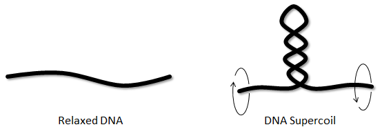
Figure 1. A coiled loop structure (plectoneme) can be formed when the DNA is twisted.
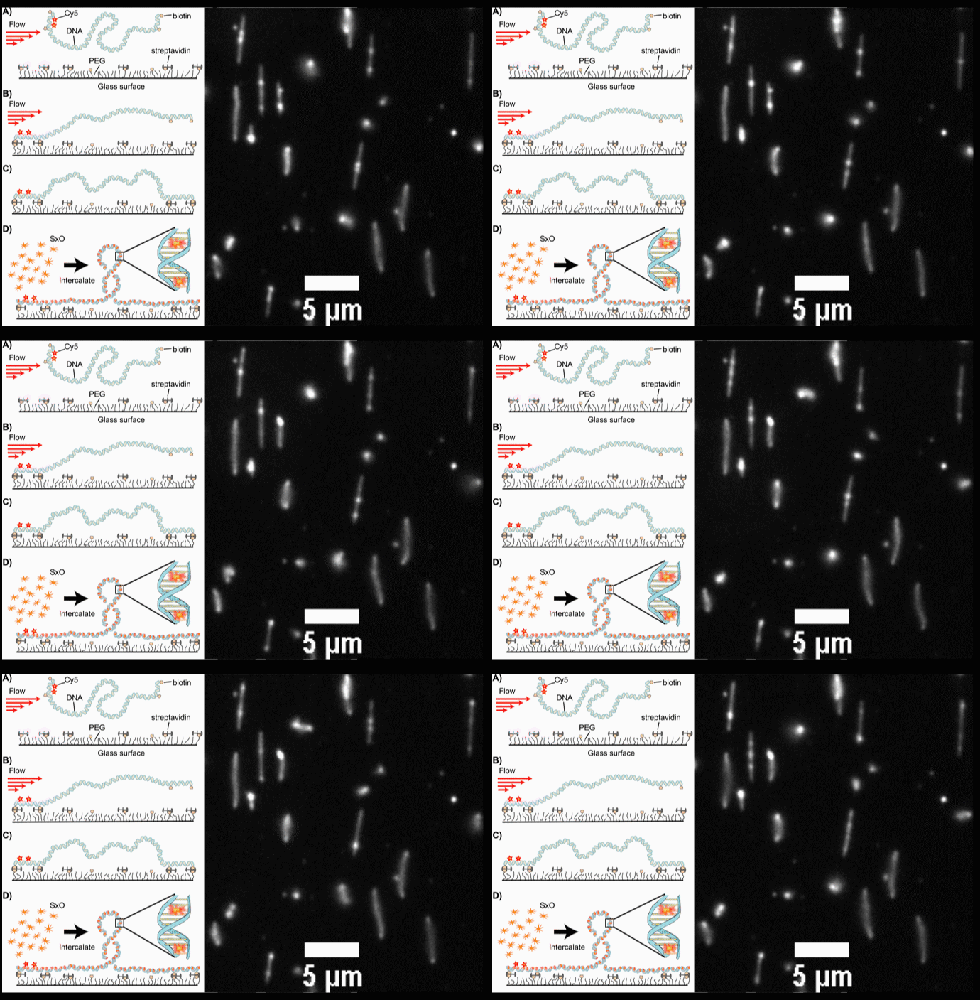
Fig. 2. (A-D) Schematic diagrams of the intercalation-induced Supercoiled DNA (ISD). (E) A fluorescence microscopy movie with multiple DNA molecules showing plectonemes as diffusing bright spots along DNA.
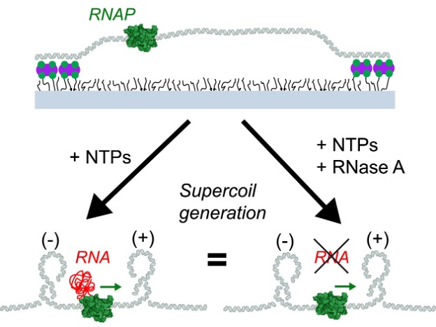
Fig. 3: Schematic of RNA polymerase –induced DNA supercoil formation regardless of viscuous drag induced by the RNA transcript.
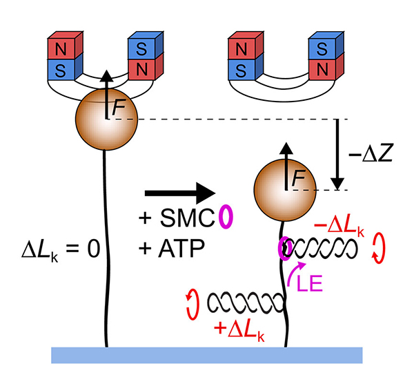
Fig. 4: Magnetic tweezer experiment show that SMC complexes induce negative supercoils into their extruded loop and negative supercoils outside of the loop.