SMC motor proteins for DNA loop extrusion
In a human cell, approximately 2 meters of DNA are compacted into micrometer-sized chromosomes. When cells divide, DNA is distributed equally into daughter cells, such that each of them inherits exactly one copy of the genome. It remains a mystery how DNA is packaged within confines of cells and equally divided when cells divide.
Recently, our group (in collaboration with Christian Haering, EMBL Heidelberg) elucidated the underlying mechanism of DNA organisation: a process called ‘loop extrusion’ that condenses DNA by progressively pulling DNA into many loops by the ring-shaped protein complex condensin, a a member of the SMC (structural maintenance of chromatin) protein complexes.
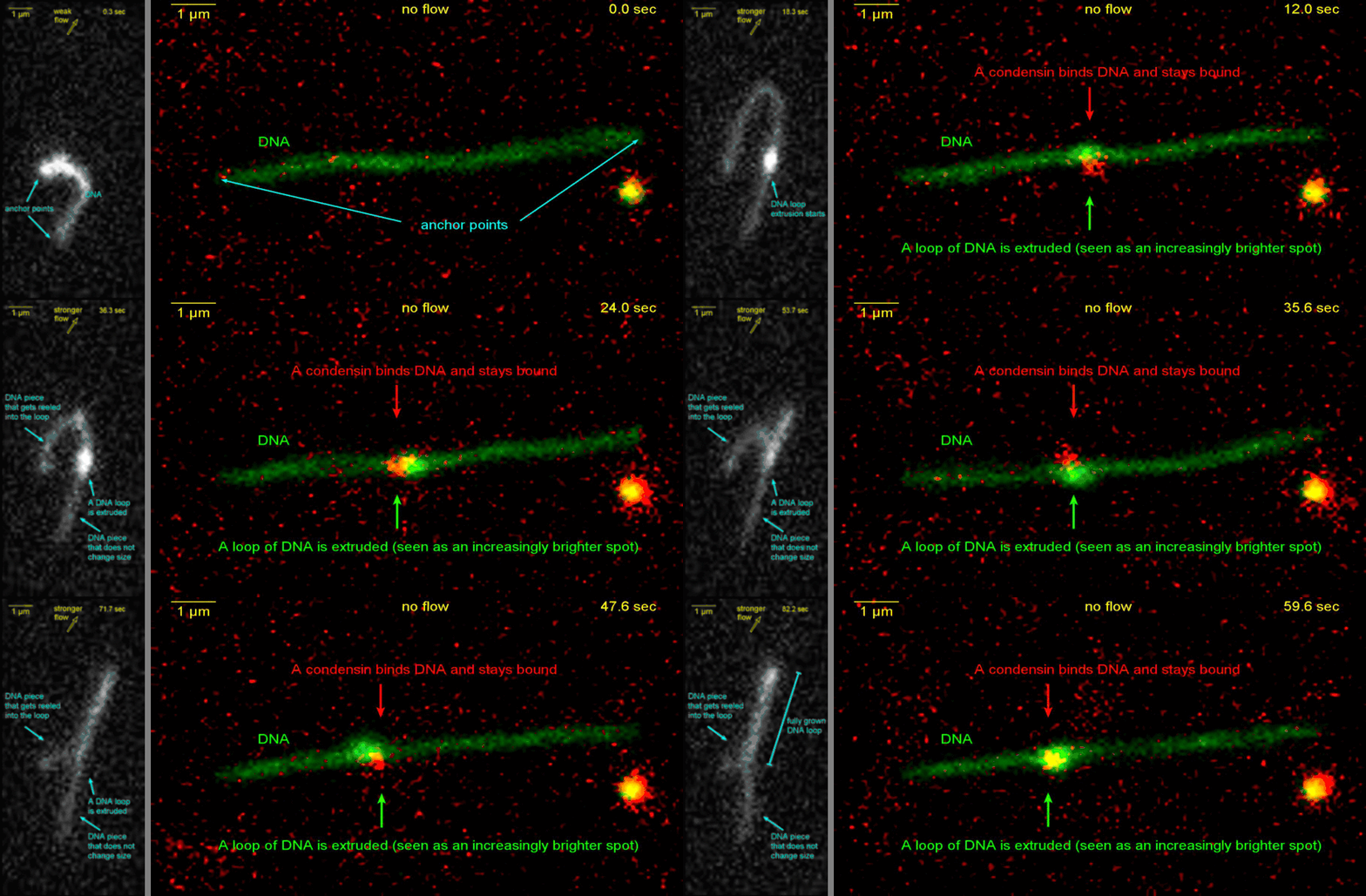
Fig 1: Real-time observation of DNA loop extrusion by condensin. (Ganji et al. 2018, Science)
Loop extrusion by SMCs: from bare DNA towards chromosome organization
The SMC (Structural maintenance of chromosomes) protein complexes form a specific class of motor protein complexes that organize DNA in nearly all living organisms. We employ various single-molecule assays like magnetic tweezers, fluorescence microscopy, and atomic force microscopy to elucidate the mechanical properties of these SMC proteins. We have shown that condensin compacts DNA upon ATP hydrolysis and the underlying mechanism of DNA compaction is by extruding loops on DNA in a progressive manner (Fig.1).
The dynamic visualization of the loop formation flooded us with details like the velocity and step size of the condensin motor, asymmetric nature of the loop extrusion, and the size of the loops; which disclosed the underlying mechanisms of condensin.
Recent works from our lab show that eukaryotic SMC complexes such as cohesin, condensin and SMC5/6 extrude DNA asymmetrically with a twist of -0.6 at each step. Further findings also show that cohesin can switch directions while loop extruding via NIPBL exchange (Fig. 2).
However, this loop extrusion on a bare DNA answer only a part of the whole puzzle of 3D organization of chromosome
We are interested in answering many other parts of the puzzle like:
i) How are human SMC complexes regulated and how do they interact with each other?
Different SMC complexes like cohesin and condensin are functionally regulated at various cell cycle stages. At certain cell-cycle phases, cohesin and condensin coexist on DNA and interact with each other. We study how these complexes influence each other and coordinate DNA looping; whether they bypass each other or stall upon encounter.
We also study how different protein regulators of hCondensin and hCohesin affect their activity and loop dynamics (such as initiation, extrusion rate, loop size and lifetime).
ii) What happens when DNA loops encounter other objects on the DNA?
In cells, SMC motors extrude loops on busy chromatinised DNA where they encounter DNA-bound obstacles, other proteins or motors of transcription and DNA replication machineries (Fig 4). We are interested in addressing what happens when SMC motors interact with such DNA bound obstacles, which would further hint at the biophysical mechanism of loop extrusion.
iii) How do extruded loops regulate other important cellular processes like gene regulation and DNA damage repair?
We try to answer these questions by single-molecule techniques like magnetic tweezers and fluorescence microscopy which helps us explore the action of SMC motors with unprecedented details.
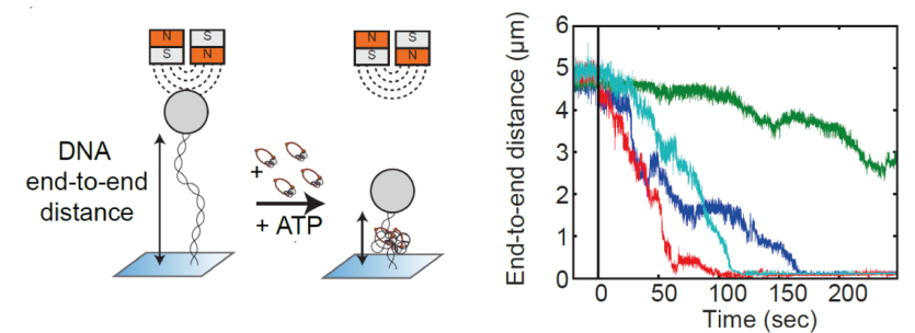
Fig.2. Real-time observation of condensin-driven DNA compaction upon ATP hydrolysis.
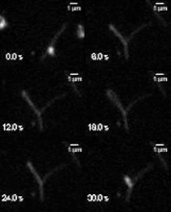
Movie 1. Visualization of a DNA loop that is extruded by condensin.

Fig.3. DNA loop extrusion direction switches coincide with an exchange of NIPBL-ΔN

Direct visualization of conformational dynamics of SMC proteins
We are studying the structure and molecular mechanism of SMC proteins using atomic force microscopy (AFM) and Förster Resonance Energy Transfer (FRET). These techniques allow us to directly relate the complex structural arrangements of SMC complexes to their function in loop extrusion.
We already succeeded to obtain a movie of dynamical conformational changes of condensin using a state-of-the-art variant of AFM, high-speed AFM. This is an excellent technique for understanding protein structure and function because of its high spatio-temporal resolution in the native liquid phase. We already demonstrated that the SMC arms are flexible and dynamic.
We are now expanding this line of research by generating FRET probes on SMC complexes to relate their loop extrusion activity directly to protein conformations. These two approaches will allow us to resolve the mechanism of loop extrusion at an unprecedented resolution.
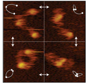
Fig 5. Frames of a high-speed AFM movie of condensin’s SMC2/4 dimer. The dynamical conformational changes show that the coiled-coils of SMC dimers are flexible and show extensive fluctuations in time (Eeftens et al. 2016, Cell Reports).

Movie 2. HS AFM video images of condensin with all subunits.